


Reorder contact lenses in just one click
Place an order with your account today, and reorder your contact lenses in just one click next time.
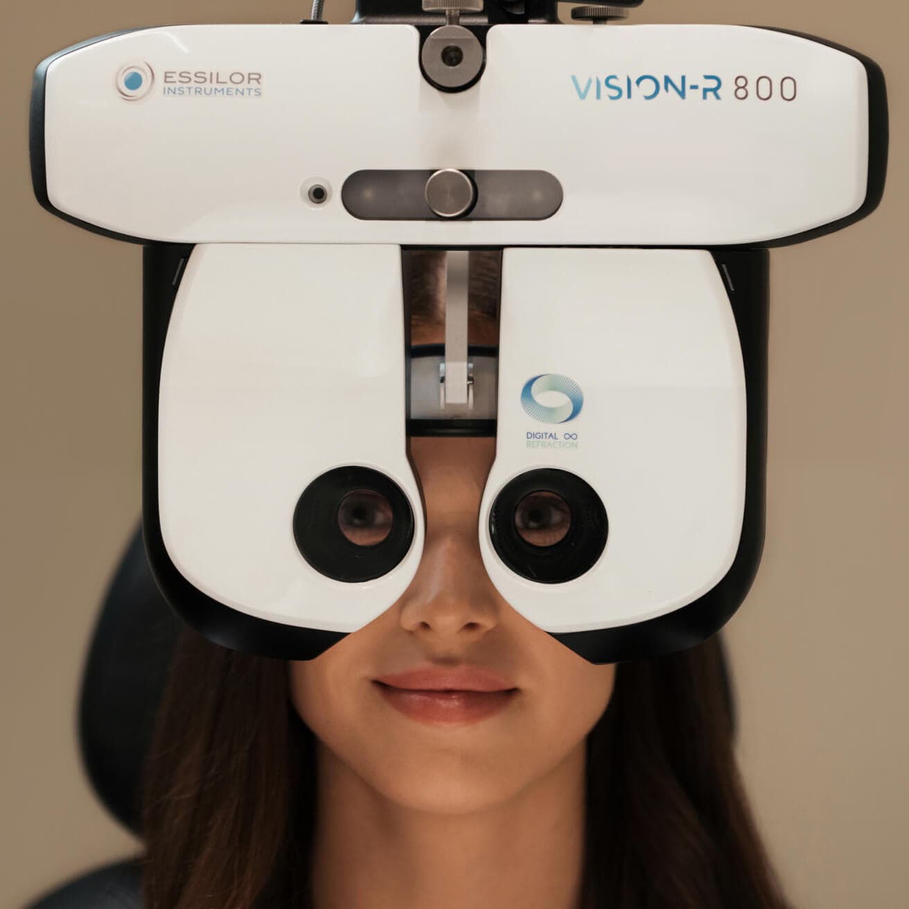
In many ways, the eye works similarly to a camera. The different parts of the eye work together to capture an image, then send it to the brain to make sense of it. When something is off — even by a little bit — it can affect how the eye functions and how well you see.

20/20 is a measurement of visual acuity, which is how sharp your eyesight is. When measuring
distance visual acuity in the U.S., the first number will always be 20. The second number will
change depending on how good or poor your vision is. The higher the second number, the blurrier
your vision is.
For example, if someone’s vision is 20/40, it means they need to stand
20 feet away to read something that a person with “normal” vision could read from 40 feet away.
A person whose visual acuity can’t be corrected better than 20/200 is considered legally
blind.
The term “20/20 vision” is used to describe vision that is average, or right
where it should be. When a patient has poor vision, the eye doctor’s goal is usually to correct
it to 20/20 with glasses or contact lenses.
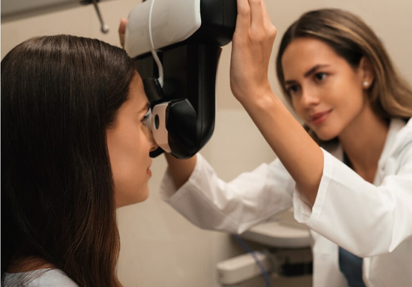
Book an eye exam in 3 easy steps
Choose your location
Schedule an eye exam
Add to calendar